Microdiscectomy is a common surgical technique for treating herniated discs in the lumbar spine.
While this procedure is generally successful, some patients may experience recurrent lumbar disc herniation, known as reherniation, which can lead to significant discomfort and require additional treatment.
Understanding the signs of reherniation after microdiscectomy is crucial for early diagnosis and effective management.
This comprehensive guide explores the symptoms, risk factors, diagnostic methods, and treatment options for recurrent disc herniation.
Understanding Disc Herniation and Microdiscectomy
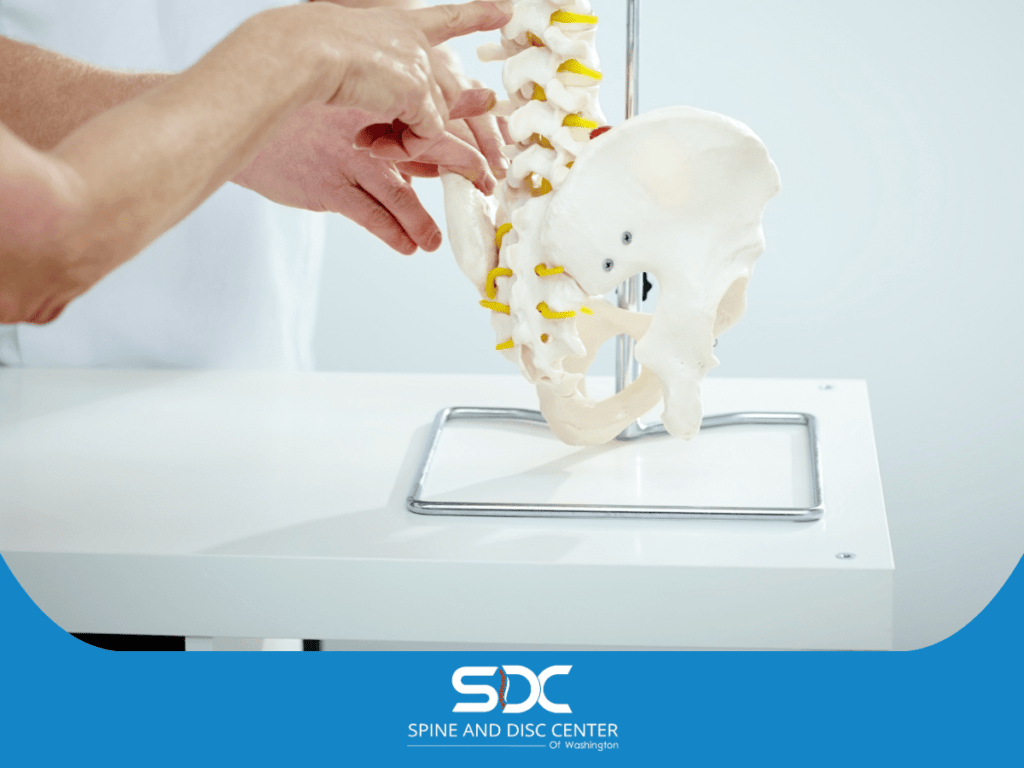
A herniated disc occurs when the spinal disc, which acts as a cushion between the vertebrae, protrudes or ruptures, pressing on nearby nerves. This condition can cause back pain, leg pain, and other neurological symptoms.
Microdiscectomy is a minimally invasive surgical technique designed to remove the herniated portion of the disc, relieving pressure on the affected nerves and alleviating pain.
The Process of Microdiscectomy
During a microdiscectomy, a type of discectomy surgery, a surgeon makes a small incision in the lower back to access the lumbar spine. Using specialized instruments and a microscope, the surgeon removes the herniated disc material, decompressing the nerve roots.
This procedure is typically performed under general anesthesia and requires a short hospital stay, with most patients experiencing significant pain relief shortly after the surgery.
Benefits of Microdiscectomy
Microdiscectomy offers several benefits, including:
- Minimally Invasive Technique: Reduced tissue damage and quicker recovery.
- Effective Pain Relief: Alleviates nerve compression, reducing back and leg pain.
- High Success Rate: Many patients report significant improvement in symptoms.
However, as with any surgical procedure, microdiscectomy carries risks, including infection, bleeding, and the potential for recurrent disc herniation.
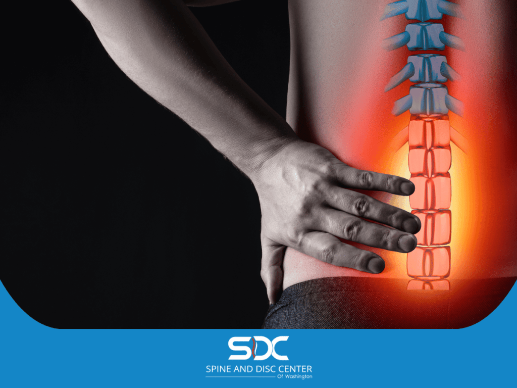
Signs of Reherniation After Microdiscectomy
Reherniation occurs when the remaining disc material protrudes again, causing similar symptoms to the initial herniation. Recognizing the signs of reherniation is vital for timely intervention.
Common symptoms include:
- Recurrent Back Pain: Patients may experience a return of back pain similar to that felt before the initial surgery.
- Leg Pain: Pain radiating down the leg (sciatica) is a classic symptom of disc herniation.
- Neurological Deficits: Numbness, tingling, or weakness in the lower extremities can indicate nerve compression.
- Decreased Disc Height: Imaging studies may show a reduction in the height of the intervertebral disc space.
- Pain During Physical Activity: Activities that stress the lumbar spine may exacerbate pain.
These symptoms can vary in severity and may develop gradually or suddenly during the postoperative period.
Risk Factors for Reherniation
Several factors can increase the risk of recurrent disc herniation after lumbar discectomy. Understanding these risk factors can help in managing and potentially preventing reherniation:
- Disc Degeneration: Patients with advanced disc degeneration are at a higher risk for reherniation.
- Body Mass Index (BMI): Higher BMI can increase the stress on the lumbar spine, contributing to disc problems.
- Poor Posture: Inadequate posture and improper lifting techniques can strain the spine and lead to reherniation.
- Primary Disc Prolapse Severity: The extent of the initial herniation can influence the likelihood of recurrence.
- Surgical Technique: The skill and technique of the surgeon play a significant role in the success of the surgery.
- Physical Activity Level: High levels of physical activity or returning to strenuous activities too soon can increase the risk.
- Scar Tissue Formation: Epidural fibrosis, or the formation of scar tissue around the nerve roots, can contribute to nerve root tethering and recurrent herniation.
Diagnostic Methods for Reherniation
Diagnosing recurrent disc herniation involves a combination of patient history, physical examination, and imaging studies. Key diagnostic methods include:
- Physical Examination: A thorough physical exam can reveal signs of nerve compression, such as weakness, numbness, or reflex changes in the lower extremities.
- Imaging Studies: MRI is the gold standard for diagnosing disc herniation. It provides detailed images of the intervertebral discs, spinal canal, and nerve roots.
- Diagnostic Nerve Root Injection: This procedure involves injecting a local anesthetic near the affected nerve root to confirm the source of pain.
- Epidural Steroid Injection: Used both for diagnosis and treatment, this injection can reduce inflammation and pain, helping to identify the involved nerve roots.
These diagnostic methods help determine the presence and extent of recurrent herniation, guiding treatment decisions.
Treatment Options for Recurrent Lumbar Disc Herniation
Treatment for recurrent disc herniation depends on the severity of symptoms and the impact on the patient’s quality of life. Options include:
- Conservative Management: Initial treatment may involve pain medications, physical therapy, and epidural steroid injections to manage pain and inflammation.
- Physical Therapy: Targeted exercises and therapies can strengthen the back muscles, improve flexibility, and reduce the risk of further injury.
- Repeat Microdiscectomy: For patients with significant symptoms, repeat microdiscectomy may be necessary to remove the recurrent disc material and decompress the nerve roots.
- Annular Closure Device: This device can be used during surgery to close the annular defect and reduce the risk of reherniation.
- Surgical Management: In cases where conservative management fails, additional surgical options such as lumbar fusion or artificial disc replacement may be considered.
Preventing Reherniation
Preventing reherniation involves adopting lifestyle changes and following postoperative guidelines to minimize stress on the lumbar spine. Strategies include:
- Maintaining a Healthy Weight: Reducing body mass index (BMI) can decrease the load on the lumbar spine.
- Proper Posture: Practicing good posture and ergonomics can reduce strain on the spine.
- Regular Exercise: Engaging in low-impact exercises such as walking, swimming, and yoga can strengthen the back and improve flexibility.
- Avoiding High-Risk Activities: Limiting activities that involve heavy lifting or repetitive bending can prevent strain on the lumbar spine.
- Following Postoperative Instructions: Adhering to the surgeon’s postoperative guidelines is crucial for recovery and preventing reherniation.
Long-Term Outlook and Patient Satisfaction
The long-term outlook for patients undergoing microdiscectomy and experiencing reherniation varies. Many patients achieve significant pain relief and improved function with appropriate treatment. Factors influencing the long-term outlook include:
- Extent of Reherniation: The severity of recurrent herniation and its impact on nerve roots and spinal structures.
- Treatment Success: The effectiveness of the chosen treatment approach, whether conservative or surgical.
- Patient Compliance: Adherence to postoperative guidelines and lifestyle modifications.
- Overall Health: The patient’s general health and presence of other medical conditions.
Patient satisfaction is generally high among those who experience successful treatment for reherniation.
Ongoing research and advancements in surgical techniques continue to improve outcomes and reduce recurrence rates.
Postoperative Care and Rehabilitation
Postoperative care and rehabilitation are critical components of recovery and prevention of reherniation. A comprehensive rehabilitation program typically includes:
- Physical Therapy: Physical therapists design personalized exercise programs to strengthen the core and back muscles, improve flexibility, and promote proper body mechanics.
- Pain Management: Pain medications, epidural steroid injections, and other pain management techniques may be used to control postoperative pain and inflammation.
- Activity Modification: Patients are advised to avoid high-impact activities and heavy lifting during the initial recovery period. Gradual return to normal activities is encouraged based on individual progress.
- Regular Follow-Up: Scheduled follow-up visits with the surgeon to monitor healing and address any concerns promptly.
- Education: Patients are educated about proper posture, body mechanics, and techniques to prevent strain on the lumbar spine.
Emerging Techniques and Technologies
The field of spinal surgery is continuously evolving, with new techniques and technologies being developed to improve outcomes and reduce the risk of reherniation. Some emerging approaches include:
- Minimally Invasive Surgical Techniques: Advances in minimally invasive spine surgery allow for smaller incisions, reduced tissue damage, and quicker recovery times.
- Annular Closure Devices: These devices are used to close the defect in the annulus fibrosus after discectomy, reducing the risk of recurrent herniation.
- Biologics and Tissue Engineering: Research is ongoing into the use of biologics and tissue engineering to promote disc regeneration and repair.
- Artificial Disc Replacement: For patients with severe disc degeneration, artificial disc replacement offers an alternative to spinal fusion, preserving motion at the affected segment.
Patient Education and Support
Educating patients about their condition, treatment options, and preventive measures is essential for successful outcomes.
Support groups and resources can provide valuable information and encouragement throughout the treatment and recovery process. Key aspects of patient education include:
- Understanding the Condition: Patients should be informed about the nature of disc herniation and the potential for reherniation.
- Treatment Options: Clear explanations of both conservative and surgical treatment options, including risks and benefits.
- Postoperative Care: Detailed instructions on postoperative care, activity restrictions, and rehabilitation exercises.
- Lifestyle Modifications: Guidance on maintaining a healthy weight, practicing proper posture, and incorporating regular exercise into daily routines.
Conclusion
Reherniation after microdiscectomy is a challenging condition that requires careful diagnosis and management. Recognizing the signs of reherniation, understanding the risk factors, and exploring the available treatment options are crucial for improving patient outcomes.
By adopting preventive measures and adhering to postoperative guidelines, patients can reduce the risk of recurrent herniation and enjoy a better quality of life.
Final Thoughts
This blog post is intended for informational purposes only and should not be blindly followed. It is not a substitute for professional medical advice, diagnosis, or treatment.
Please schedule an appointment with our qualified healthcare provider for personalized diagnostic and guidance tailored to your specific condition.